6 Neonatal Emergencies
6.1 Introduction
The usual newborn cries on delivery, is pink all over, well flexed and moves all limbs spontaneously, suckles well at the breast, breaths normally and weighs between 2.5 – 4.0kg. Neonatal emergencies are not uncommon and encompass a wide range of conditions occurring in the first 28 days of life. The classical mnemonic for these is THE MISFITS.
Trauma/Abuse, Heart & Lung, Endocrine, Metabolic disturbances, Inborn errors of metabolism, Sepsis, Formula, Intestinal, Toxins, Trisomies, and Seizures.
6.2 Approach to newborn emergencies
Presenting features of many serious neonatal disorders are nonspecific. The history and physical examination are essential in the overall approach to the patient. Prenatal, perinatal and postnatal history play a huge role in neonatal assessments They guide and inform the health worker on the most appropriate investigations, which would eventually lead to a correct diagnosis. A complete history may unmask the likely cause of symptoms and guide further questioning, for example, sepsis.
Examination on the other hand involves assessing the Airway, Breathing, Circulation, Random Blood Sugar, provision of Oxygen and checking Oxygen saturation. This can be pre- and post-ductal. Others include temperature checks and other vital signs. The weight, current weight, head circumference, and length. Intravenous access should be obtained for possible further treatment. Appropriate investigations should also be done.
Requisite investigations should also be done accordingly.
6.3 Respiratory Emergencies
This is one of the most common and includes:
- Primary pulmonary Hypertension,
- Meconium Aspiration Syndrome,
- Congenital Pneumonia
- Birth Asphyxia and
- Respiratory Distress Syndrome
6.3.1 Respiratory Distress Syndrome
RDS is due mainly to a lack of surfactant in the lungs. Surfactants are essential for reducing the surface area of the lungs, thus helping in breathing. Incidence and severity increase with decreasing gestational age. Other risk factors include prematurity, male gender, multiple gestations, being born to a mother with diabetes mellitus and hypothermia. Signs of RDS include tachypnea, grunting, recessions and cyanosis. Prevention involves preventing preterm births and administering corticosteroids to the mother of gestation between 24 to 34 weeks before delivery. Treatment however involves the administration of surfactant.
Note that this condition is different from Respiratory Distress in a newborn. Respiratory distress is a more generalised term used as a single or a combination of signs as a result of increased work of breathing. It can result from pulmonary as well as non-pulmonary causes. These include cardiac, neurological (eg. Asphyxia), haematological (Anemia) and sepsis. It occurs in both term and preterm children.
6.4 Endocrine Emergencies
Neonatal jaundice is the most prominent endocrine disorder in this section. This is appropriately discussed in Section 7.1
6.5 Metabolic Emergencies
6.5.1 Hypoglycemia
Hypoglycemia is common in the stressed neonate and glucose levels should be monitored regularly. In the newborn period, it is defined as a random blood glucose of < 2.6mmol/l. Risk factors include sepsis, Infant of a diabetic mother, prematurity, Intrauterine growth restriction, birth asphyxia and hypothermia. Signs include Lethargy, poor feeding, seizures, and apnea. Neurological damage may result from hypoglycemia in neonates.
Neonatal hypoglycemia is most commonly seen in macrosomic infants and infants of diabetic mothers. For these babies, during pregnancy, maternal glucose crosses the placenta to cause fetal hyperglycaemia. The fetal pancreas responds by increasing insulin production. Following delivery, the hyperglycaemic stimulus is instantly removed but insulin production may take longer to slow down. This results in an increased risk and incidence of hypoglycemia at the early newborn period.
Management involves initially checking the airway, breathing and circulation. 2ml/kg of IV 10% Dextrose or 5ml/kg of 5% Dextrose may be given PR if IV access is unavailable. Ensure the baby has a normal body temperature (temperature target: 36.50 – 37.50C) as hypothermia prone the baby to hypoglycaemia. Always look for the underlying cause of the hypoglycemia and treat it appropriately.
6.6 Neonatal Sepsis
Sepsis in the neonate kills more than a million babies worldwide every year. It is a clinical syndrome characterized by signs of infection with accompanying bacteremia in the first month of life. It can be categorized into Early Onset Neonatal Sepsis (EOS), which refers to the presence of signs of infection accompanied by a positive culture within the first 72 hours of life, and Late Onset Neonatal Sepsis (LOS), which signifies the onset of signs of infection with a positive culture after 72 hours of life.
Causative organisms: Early-onset sepsis is typically caused by organisms from the maternal genital tract, whereas late-onset sepsis is caused by organisms in the caregiving environment or community. Common organisms are Klebsiella pneumoniae, E. coli, and Coagulase Negative Staphylococcus among others. For many of these organisms, the resistance rate to antibiotics is alarmingly going up.
Antenal risk factors known to be associated with neonatal jaundice include spontaneous rupture of membranes, less than 37 completed weeks of gestation, spontaneous preterm labour, rupture of membranes greater than 18 hours before delivery, maternal chorioamnionitis, maternal fever of 38 degrees Celsius or more, maternal invasive bacterial infection requiring antibiotics, pre-labour rupture of membranes, Group B Streptococcus infection in a previous baby, or current pregnancy, meconium-stained amniotic fluid and foul-smelling liquor.
Postnatally, risk factors for neonatal sepsis include prolonged resuscitation at birth, prematurity, invasive procedures, mechanical ventilation, excessive handling, home delivery, lack of hand washing and inadequate Infection prevention control. Others include overcrowding and prolonged hospital stay.
Signs of neonatal sepsis include
- Abnormal colour: Pale, cyanotic, mottled appearance, jaundice, grey
- Temperature instability
- Abdominal signs: distension, poor feeding, vomiting, diarrhoea
- Respiratory: Apnea, respiratory distress, gasping (Abnormal breathing)
- Hypo - or hyperglycemia
- Cardiovascular: Shock, tachycardia (HR > 180), Bradycardia (HR < 80)
- Abnormal bleeding
- Central Nervous System: Excessive crying, irritability, seizures, altered tone, lethargy,
Management of neonatal sepsis should be comprehensive. It should include an initial evaluation for resuscitation of the airway, breathing and circulation. Further, the blood sugar should be measured. Early reversal of the shock state by administering an initial bolus of 10ml/kg of crystalloid or its equivalent should be done in the shock present. Vasopressors or inotropes should be used in septic shock only after appropriate volume resuscitation has been done. The goals of the resuscitation should be Normal Cap refill (less than 2 seconds), normal pulses, warm extremities, and appropriate urine output (greater than 1mL/kg/hr).
Increased successful treatment of neonatal sepsis requires early recognition and urgent administration of appropriate antibiotics.
After resuscitation, ongoing management usually starts with a detailed history to assess risk factors and other presentations. A thorough physical examination will then be performed, looking for and documenting specific signs indicating severity.
Investigations usually include a blood culture. This is considered the gold standard for diagnosis. Ideally, a culture should always be done before the first dose of antibiotics. Other auxiliary investigations include a complete blood count, blood gases, urine culture, and a lumbar puncture. The threshold for performing a lumber puncture in all symptomatic newborns suspected of sepsis should be encouraged. It should however be deferred in neonates considered too unstable to tolerate the procedure, or where there is an absolute contraindication.
Supportive treatment is essential for a good outcome in neonatal sepsis. Antibiotics are not the entire solution to their treatment. Nutrition or breastfeeding should be optimized. The environment should be thermo-neutral and oxygen saturation should be maintained within the normal range (89 – 95%). Intravenous fluids should be used if the infant is hemodynamically unstable. Monitoring of the blood glucose levels should be instituted. Packed red cells and fresh frozen plasma should be used in the event of anaemia or bleeding.
6.7 Gastrointestinal emergencies
Gastrointestinal emergencies in newborns can be broadly divided into the following:
Obstructive: Some of these are Tracheoesophageal fistula, Duodenal Atresia, Hirschsprung’s Disease, Biliary Atresia, Pyloric Stenosis, Intestinal Volvulus, Imperforate Anus and Necrotizing enterocolitis.
Abdominal wall defect: The most notable examples here are Omphalocele and Gastroschisis
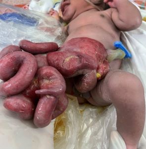
6.7.1 Gastroschisis
This is an anterior abdominal wall defect, located to the right of the umbilicus, and contains herniated intestines that have no material covering the sac. It occurs in approximately 1 in 10000 births. Rarely, it is associated with other genetic syndromes. However, it may be associated with intestinal atresia, stenosis and malrotation. Other associations include prematurity (50-60%)and cryptochidism (31%). Generally, it has a better prognosis compared to an omphalocele. The prognosis is excellent for small defects. Mortality is expertise and facility-dependent but generally around 5 to 10%. Necrotizing enterocolitis is a well-recognised complication, occurring in as much as 18%.
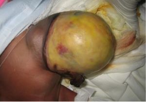
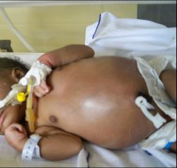
6.7.2 Omphalocele