26 Tetralogy of Fallot
26.1 Definition
Tetralogy of Fallot (ToF) is a conotruncal defect resulting from anterior misalignment of the infundibular septum, giving rise to four components:
- Large, nonrestrictive Ventricular Septal Defect
- The aorta overriding the interventricular septum
- Right ventricular outflow tract obstruction, including at the infundibulum, main and branch pulmonary arteries
- Right ventricular hypertrophy
26.2 Incidence/prevalence
It is the most common cyanotic congenital heart disease. About 1 in 3500 babies born in the US are born with ToF. Accounts for 7–10% of all congenital cardiac malformations. More common in Males. In a series of echocardiograms done in Kumasi, Ghana, ToF was the most common cyanotic congenital heart disease seen in 13% of all heart diseases. There was a significant male preponderance with a female vs male prevalence of 10% vs 15%, respectively.
26.3 Aetiology
Although there is no known definite cause as to why some babies develop ToF in utero, certain environmental and biological factors are known to increase the risk:
Genetic: CHARGE syndrome, chromosome 22q11 microdeletion (Di George syndrome), Down syndrome, Edward’s syndrome, or Patau syndrome, VACTERL association
Teratogens: maternal diabetes mellitus, retinoic acid exposure, maternal phenylketonuria (PKU), Alcohol (fetal alcohol syndrome), Warfarin (fetal warfarin syndrome), Trimethadione: antiepileptic drug
26.4 Pathophysiology
- The severity of clinical signs and symptoms depends on the proportion of the cardiac output going through the pulmonary artery, the relative pressures in the right and left ventricles, and the proportion of the aorta overriding the VSD.
- The VSD is normally of a significant size, which causes the systolic pressures between the ventricles to equalize. In mild ToF, the left ventricular pressures remain higher than the right ventricle, thus, blood shunts from left to right through the VSD. These patients are normally acyanotic. In more severe diseases, due to increased right ventricular pressure (secondary to Pulmonary Stenosis), the shunt direction reverses from right to left, allowing the mixing of deoxygenated and oxygenated blood. This results in lower oxygenated blood in the systemic circulation, making patients cyanotic.
- Pulmonary stenosis can be classified according to its location. The commonest site is the infundibular septum (50%). The stenosis may also be valvular (10%) or a combination (30%). This results in impaired flow of deoxygenated blood into the main pulmonary artery. It may be severe enough to cause intermittent right ventricular outflow tract obstruction. This forms the basis of hypercyanotic episodes (tet spells).
- Hypertrophy of the right ventricle occurs in response to the high pressures it must overcome to force deoxygenated blood through the right ventricular outflow tract obstruction. Compared to the normal heart, the aorta in ToF is dilated and displaced over the interventricular septum. Aortic dilatation is caused by an increase in blood flow through the aorta as it receives blood from both ventricles via the Ventricular Septal Defect.
26.5 Signs and symptoms
Patients with Tof are often not born with cyanosis. This may lead to the diagnosis being missed at birth. However, progressive cyanosis occurs in the first year of life. They usually have little or no signs of heart failure. Most will present with poor exercise tolerance, which worsens as the child ages. In children who can walk, frequent squatting is observed in unrepaired ToF. Other features include poor feeding and poor weight gain.
Physical examination of children with ToF may reveal digital clubbing of varying stages, central cyanosis, and plethora, usually seen in the hands and eyes. Auscultation classically reveals the first and second heart sounds with an ejection systolic murmur loudest at the upper to middle left sternal edge.
26.6 Investigations
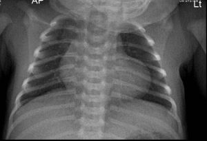
Bedside pulse oximetry often reveals an oxygen saturation of less than 90%. A chest X-ray shows a normal-sized heart with a classical boot shape and decreased pulmonary vascular markings. In about 30% of the cases, a right arch is present.
An electrocardiogram, though non-specific, may show right axis deviation, right ventricular hypertrophy, and right atrial enlargement.
An echocardiogram is the most useful diagnostic modality. It delineates the defect by showing the anterior malalignment Ventricular Septal Defect, degree of infundibular stenosis, state of the pulmonary artery and/or branches, and the overriding aortic arch sidedness. Other anatomical abnormalities that may co-exist, eg, Atrioventricular Canal Defects, Atrial Septal Defect, and coronary artery abnormalities can also be obtained. A CT angiogram and Magnetic Resonance Angiography can be done in complex cases and in preparation for surgery.
26.7 Treatment
Since patients with ToF hardly experience heart failure, antifailure medications are not the mainstay of treatment.
Untreated cyanotic congenital heart disease is associated with a chronic hypoxic state, which leads to polycythemia. This leads to a high demand for iron and increases susceptibility to iron deficiency. Furthermore, it is well documented that iron deficiency increases the chance of a hypercyanotic spell and stroke. Iron treatment in ToF is therefore key in its outpatient management. Nutritional rehabilitation is done for chronically malnourished patients.
Surgical therapy is the definitive treatment. A Blalock-Taussig shunt can be done to improve oxygenation. A stand can also be inserted in younger children when deemed necessary. Definitive corrective surgery should be done for all children with ToF.
26.8 Natural history
Features of ToF are progressive if not corrected surgically. There is usually progressive dyspnoea on exertion and cyanosis as the child ages. However, some “pink tets” can live very well into adulthood. Untreated, approximately 50% will live to their 6th birthday.
26.9 Complications
A feared presentation in untreated children with a ToF is the hypercyanotic spell (Tet spell). This is treated further below. Other complications can result from the embolus effect, leading to stroke and cerebral abscess. High hematocrit may lead to hyperviscosity, headache, and dizziness. Infective endocarditis is another known complication. Long-term complications include right ventricular dysfunction, coagulopathy, and arrhythmias.
26.10 Prognosis
Repaired, the 25-year survival is about 95%.(Smith et al. 2019) Unrepaired, most will die by their 10th birthday. This is especially so for those with other genetic syndromes and associated malformations.
26.11 Differential diagnosis
Cyanosed ToF patients have a differential diagnosis of Transposition of the great arteries, Tricuspid atresia, pulmonary atresia, etc. Pink ToFs will have a differential diagnosis of a Ventricular Septal Defect
26.12 Prevention
There is no known prevention for ToF. However, it is always prudent for prospective mothers and those in the first trimester to avoid recreational drugs, alcohol, and some over-the-counter medications. Also, folic acid supplementation should be encouraged.
26.13 Hypercyanotic spell
A hypercyanotic spell (tet spell) is an emergency in children with ToF and, to a lesser extent, other cyanotic congenital heart diseases. It presents most commonly in children less than 2 years old.
26.13.1 Presentation
Children with hypercyanotic spells present with paroxysms of increased and deep breathing, irritability, prolonged, unsettled crying, increasing cyanosis, seizures, and decreased intensity of the heart murmur. In untreated cases, this might lead to brain damage or death.
26.13.2 Pathophysiology
A hypercyanitic spell can have many precipitating factors. These may include fever, anemia, dehydration, prolonged crying, and eating. These precipitants lead to decreased lung blood flow and progressively increase right-to-left shunting. This leads to increasing cyanosis, tachycardia, and reduced systemic vascular resistance. Carbon dioxide accumulation stimulates the central respiratory center, leading to increased and deep breathing. All these unfortunately cause further right-to-left shunting, thus perpetuating the hypoxia.
26.13.3 Treatment
The treatment goal is to increase preload and promote pulmonary blood flow.
- First, the child should be placed in a knee-chest position. This increases systemic vascular resistance, temporarily raises the systemic pressure, and reduces the right-to-left shunting.
- Oxygen can be administered, though it is of limited value and should not be forced on the patient if he is combative.
- Next volume expansion with intravenous fluids should be administered. This raises the preload and increases systemic pressure, thus reducing right-to-left shunting.
- Intramuscular or subcutaneous morphine can be administered. This aids in relaxing the infundibular muscle and thus promotes pulmonary blood flow. It also sedates the child, thus making him/her less acidotic. Acidosis perpetuates the hyperchaotic spell
- Intravenous propranolol, esmolol, or metoprolol is administered to reduce the right ventricular outflow tract obstruction.
- Some alpha-agonists, such as Phenylephrine, can be given to improve blood pressure in severe cases
- Long-term treatment may include oral propranolol for prophylaxis, iron supplementation, and surgical correction.