5 Neonatal Delivery Pathologies
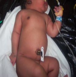
5.1 The health newborn
- Cries / Breathes normally
- Pink all over
- Well-flexed & moves all limbs spontaneously
- Suckles well at the breast
- Birth weight 2.5 – 4.0kg
- Normal vitals signs
5.2 Occurrences at birth
- The fluid in the alveoli is absorbed and replaced by air. If the transition is not smooth, it results in insufficient oxygen delivery to the vital organs…
- Poor muscle tone
- Respiratory distress or depression
- Slow heart rate
- Low BP
- Cyanosis
5.3 Birth Asphyxia
5.3.1 Definition
Birth Asphyxia is the medical condition resulting from deprivation of oxygen in the newborn that lasts long enough during the birth process to cause harm, usually to the brain.
5.3.2 Risk factors
Any condition that will lead to impairment of oxygenation or blood flow to the newborn’s brain in the perinatal period. These include:
- Prolonged labour (CPD)
- Placental failure
- Cord around the neck
- Problems with oxygenation of maternal blood / maternal disease
- Anaemia and bleeding in the baby
- Congenital heart disease
- Infections
- Deficient medical skills and or knowledge
5.3.3 Presentation
The asphyxiated baby may have any of the following:
- Poor Apgar Scores
- May not cry at birth
- Floppy/spastic
- Breathing problems
- Unresponsive
- Seizures
- Irritable
5.3.4 The APGAR Score
It is an objective method of quantifying the newborn’s condition. And is useful for conveying information about the newborn’s overall status and response to resuscitation
| 0 | 1 | 2 | |
|---|---|---|---|
| Heart Rate | 0 | <100 | >=100 |
| Respiration | 0 | Weak or Irregular | Good Cry |
| Reaction | None | Slight | Good |
| Colour | Blue or Pale | Pink body limbs blue |
All pink |
| Tone | Limp | Some movement | Active movement, limbs well flexed |
8-10 = No Asphyxia
5-7 = Mild Asphyxia
3-4 = Moderate Asphyxia
0-2 = Severe Asphyxia
5.3.5 Management
- Largely supportive
- Newborn resuscitation/oxygenation
- Correction of fluid & electrolyte imbalances including shock
- Control of seizures
- Treatment of any underlying infection
- Look out for birth injuries
- Active cooling found to improve neurological outcome
- Temperature maintenance
- Full Blood Count, Culture & Sensitivity, Blood glucose etc
- Serum electrolytes: Na, K, Ca & Mg
- Start empiric 1st line antibiotics according to protocol: / X’pen & Gentamycin
- Start IV Fluids at 50ml/kg (Plain 10% Dextrose).
- Pass a urethral catheter and monitor the baby’s urine output.
- The target temperature of the baby is 36.50C – 37.50C
5.3.6 Hypoxemic Ischaemic Encephalopathy
- The most important consequence of birth asphyxia is
- The outcome ranges from complete recovery to death
- 25 - 30% end up with permanent damage like Cerebral palsy & Mental retardation
- Prognosis dependent on gestational age, management of metabolic & cardiopulmonary complications & the severity of the encephalopathy
- Subsequent competent care and available facilities also influence the outcome
5.4 Birth Injuries
A birth injury can simply be referred to as any form of damage incurred by the baby during the birthing process. Injury may occur as a result of inappropriate or deficient medical skill or attention or may occur despite skilled and competent obstetric care.
Predisposing conditions include:
- Cephalopelvic disproportion (CPD) / Small maternal stature / Primiparity
- Macrosomia
- Shoulder Dystocia
- Prematurity
- Prolonged or precipitous labour
- Abnormal presentation
- Instrumentation
- Handling after delivery
5.4.1 Fracture
Generally, the affected limb looks deformed or swollen, and the baby barely moves it on account of pain
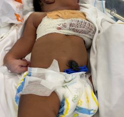
5.4.1.1 Clavicle
This is the most fractured bone during delivery; mostly during delivery of the shoulder in vertex and of the extended arms in the breech. Signs of a fracture may include no free arm movement on the affected side, crepitus and bony irregularity, and absent Moro reflex. It has an excellent prognosis, even though it is commonly missed. Treatment, if any, includes immobilization of the arm and shoulder as shown below.
This is the first type of humeral fracture
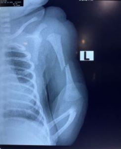
5.4.1.2 Humerus
The x-ray below shows another commonly fractured bone, the humerus.
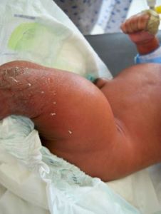
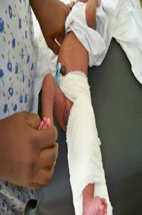
5.4.1.3 Femur
Risk factors: big baby, breech presentation, incompetency. The affected thigh looks deformed, swollen and may be reddened. The main mode of management involves splinting the limb from the waist to below the knee.
5.5 Nerve injuries
5.5.1 Brachial plexus injuries
The nerves of the brachial plexus may be compressed, stretched or torn in a difficult delivery. Paralysis occurs as a result of nerve compression from either haemorrhage or oedema. Permanent paralysis can occur from the tearing of the nerve or avulsion of the nerve root from the spinal cord or oedema. Erb’s palsy (C5-C6) is the most common type of BPI and is associated with a lack of shoulder motion. The involved extremity lies adducted, prone, and internally rotated. Grasp reflex is usually present and prognosis is generally good. Also described as the Waiter’s tip position.
5.5.2 Klumpke’s paralysis
5.5.3 Facial nerve paralysis
Loss of voluntary muscle movement in the face on account of pressure on the facial nerve during the delivery process. Risk factors include instrumental delivery, poor delivery skills, big baby etc. Usually resolves spontaneously after a few months
5.6 Scalp Injuries
5.6.1 Cephalhematoma
Tearing or disruption of the superficial veins under the periosteum leads to haemorrhage and subsequent swelling. Suture lines confine the cephalhematoma and limit the extent of the bleeding. There could be an underlying linear skull fracture. Prognosis is good with most of them resolving between 2 weeks to 3 months.
5.6.2 Subgaleal Hemorrhage
The subgaleal space is located between the galea aponeurotica & the periosteum. The space extends from the orbital ridges to the nape of the neck and laterally to the ears. Bleeding is caused by damage to the large emissary veins located in the subaponeurotic layer. The bleeding associated with subgaleal haemorrhages can be extensive. Clinically, the baby may present with pallor and lethargy, followed by tachycardia, tachypnea and hypotension. The scalp may appear tight and boggy and complications include anemia, hypovolemic shock and jaundice.
5.7 Visceral injuries
5.7.1 Liver and spleen
Usually results from pressure on the liver during delivery of the head in breech presentations. Risk factors include macrosomia, intrauterine asphyxia, extreme prematurity, and hepatomegaly
5.8 Respiratory Distress
5.8.1 Meconium aspiration syndrome
Fetuses sometimes pass meconium whilst in utero as a result of some form of stress. If the stress has been going on for a while and the fetus has been passing meconium for a few days, the cord, skin and nails may be stained. Occurs when the fetus passes meconium into the surrounding liquor and then aspirates this into the lungs. Tends to happen in term and post-date babies. Distressed fetuses tend to pass meconium either just before or during the delivery process. The smaller the amniotic fluid volume, and the more meconium the baby passes, the thicker the fluid and the more dangerous it becomes if aspirated.
History of Pregnancy and delivery looking for predisposing factors such as fetal distress, post-maturity, meconium-stained liquor etc. Physical examination looking for signs of meconium-staining on the baby, and evidence of respiratory distress (fast breathing, chest indrawing, cyanosis etc.). Investigations include FBC, Blood C&S, and sometimes a chest X-ray depending on the severity. Management is mainly supportive. Includes antibiotics, respiratory support, supportive treatment, IVFs and nutrition.
5.8.2 Transient tachypnoea of the newborn
Caused by delay in clearance of fetal lung fluid. Typically resolves within 72 hours. Often associated with Caesarean Section Delivery. Severity varies but is often mild with just tachypnoea. Management involves supportive treatment of the respiratory with oxygen.