86 Bacterial Skin Infections
86.1 Introduction
The skin is sterile at delivery and becomes colonised shortly after. The normal skin of healthy infants and children is resistant to invasion by most bacteria because the cutaneous surface serves as a dry mechanical barrier.
The transient flora consists of multiple organisms that are deposited on the skin from the environment presumably, they do not proliferate and are removed easily by washing or scrubbing the affected area.
The resident flora consists of a smaller number of organisms which are found regularly in appreciable numbers on the skin of normal individuals, multiply on the skin, form stable communities on the cutaneous surface, and are not easily dislodged.
Pathogenic bacteria, not ordinarily a regular part of this flora, persist on the skin if there is a continuous replacement from some internal or external source or if the integrity of the skin is disrupted by injury or disease.
Staphylococcus aureus forms part of the normal human flora. 10% to 30% of individuals are nasal carriers of Staphylococcus aureus and 70% to 90% are transient carriers.
86.2 Impetigo
Question:
A 4-year-old male child presents to the clinic with a one-week history of a fluid-filled eruption at the chin which easily ruptures and spreads to other areas. Mother complains his 2-year-old sister has also developed similar lesions 2 days ago.
- What is your most likely diagnosis?
- How will you manage this family?
86.2.1 Definition & incidence/prevalence
Impetigo is a common, contagious superficial skin infection. It is seen in all age groups, most common in infants and children. It can involve any body surface but occurs most often on the exposed parts of the body, especially the face, hands, neck and extremities.
86.2.2 Aetiology
It is caused by streptococci, staphylococci or both.
86.2.3 Pathogenesis
There are two classic forms
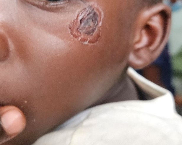
- Non-bullous impetigo – accounts for >70% of cases. It is caused by S. aureus or S. pyogenes. Spread by direct contact or through fomites. It begins with a 1 to 2mm erythematous papule or pustule that soon develops into a thin-roofed vesicle or bulla surrounded by a narrow rim of erythema. The vesicle ruptures easily with the release of a thin, cloudy, yellow fluid that dries, forming a honey-coloured crust, the hallmark of non-bullous impetigo. S. pyogenes may cause post-streptococcal glomerulonephritis if the nephrogenic strain is involved. This is shown in Figure 86.1
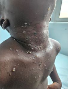
- Bullous impetigo - This is always caused by Staphylococcus aureus. Presents as flaccid, thin-walled bullae or more commonly, tender, shallow erosions surrounded by a remnant of the blister roof. High reservoir sites include nasal carriage of asymptomatic persons and perineum. This is shown in Figure 86.2
86.2.4 Investigations
Diagnosis is usually made clinically, however, aspirate of the fluid from the lesion can be sent for culture and sensitivity pattern in extensive cases.
86.2.5 Treatment
- Local skin care: cleansing, removal of crust
- Mild and localised disease- topical antibiotics e.g. Mupirocin
- In severe or more widespread diseases – oral antibiotics, e.g.: amoxicillin plus clavulanic acid, erythromycin
Note: Staph eradication can be considered in people with recurrent impetigo.
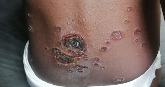
86.3 Ecthyma
Ecthyma is a deep or ulcerative type of non-bullous impetigo. Commonly seen on the lower extremities and buttocks of children. Lesions are painful and heal slowly with scar formation. The initial lesion is a vesiculopustular with an erythematous base and firmly adherent crust. Removal of the crust reveals an underlying saucer-shaped ulcer and raised margin.
86.3.1 Treatment
This is with warm compresses and an appropriate systemic antibiotic.
86.3.2 Complications
- Cellulitis
- Acute post-streptococcal glomerulonephritis in 5% of cases of non-bullous impetigo by S. pyogenes (serotypes 1, 4,12, 25 and 49)
86.3.3 Prognosis
Impetigo heals well without scarring
86.3.4 Differential diagnosis
The most common differentials to consider include insect bite, eczematous dermatoses and herpes simplex viral infection (HSV) in non-bullous impetigo and thermal burns, bullous insect bite reactions and HSV in bullous impetigo.